
Patient Info
ACL Surgery
Anterior Cruciate Ligament (ACL)
Normal Anatomy
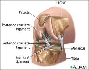
- Bones
- Femur (Thigh Bone)
- Tibia (Leg Bone)
- Patella (Knee Cap)
- Ligaments (Attach Bone to Bone)
- Anterior Cruciate Ligament (ACL)
- Posterior Cruciate Ligament (PCL)
- Medial Collateral Ligament (MCL)
- Lateral Collateral Ligament (LCL)
- Cartilage
- Menisci (Cushions Between Femur and Tibia)
- Articular Cartilage (Shock absorber on end of bone
What is the ACL?
Injury to the ACL is one of the most common sports related knee injuries.
- Location & Function
- Attaches thigh bone to the leg bone and prevents the leg bone from moving forward (towards the front) relative to the thigh
- Why does it tear?
- Usually a twisting injury or a direct blow injury to the knee
- How does a tear present?
- Sudden pain in the knee
- Often associated with a "pop"
- Swelling or fluid on the knee develops over the first hour or so after injury
- The sensation of knee instability or the bones moving over one another
- Why is it painful?
- From the swelling and injury to the joint lining which has nerve endings in it
Associated Injuries
- Meniscal Tears
- Articular Cartilage Damage
- MCL, PCL, LCL
Diagnostic Studies - Role of X-Ray, MRI
X-Rays
An x-ray is a 2 dimensional view of a bone. PLEASE BRING ALL STUDIES TO THE APPOINTMENT.
X-Rays obtained at the time of visit:
- AP OF BOTH KNEES
- AP OF BOTH KNEES WITH SOME FLEXION
- LATERAL OF INJURED KNEE
- KNEE CAP VIEW
MRI
Although the gold standard for diagnosing an ACL injury is physical exam, the MRI confirms the injury with good accuracy and also evaluates for other associated injuries.
Treatment Options
Physical Therapy
P.T. is an extremely important component to ACL injury. Regaining full motion before surgery is critical for post-operative motion.
The ACL does not have to be reconstructed in sedentary individuals, but higher level activity in a knee with no ACL puts the cartilage and other structures at risk for injury.
Will PT heal/fix my Ligament?
This is a question asked of us all the time, and the simple answer is no. However, surgery is not always necessary if return to higher level activity is not wanted, and there is no associated pain or symptomatic instability.
PRE SURGERY PT PROTOCOL
Pre surgery physical therapy is very important for those who present with extreme weakness or stiffness.
What is a ACL Reconstruction Surgery?
Arthroscopy refers to a procedure done within a joint through small incisions, using an arthroscope (camera) to visualize the structures within. Arthroscopic procedures are commonly done on an outpatient basis.
The ACL cannot be repaired; it must be reconstructed as a new ligament. This is done by using a tendon graft either from the patient's leg or from cadaver donor. The tendon is positioned in place of the injured ACL through tunnels which are drilled in the bone. The tendon is fixed in place with screws, or other fixation devices. Once the tendon has healed to the bone, these fixation devices become non-functional but do not usually need to be removed.
Incision Sites:
Two to three small dime sized incisions are made to pass the camera and instruments into the knee. One additional larger incisions of about 3-4cm is used to harvest the hamstring tendon or patellar tendon graft. These are closed with sutures that will be removed in the office in 10-14 days after surgery.
Common Grafts used for ACL surgery
- Patellar tendon
- Benefits:
- Strongest Fixation to Bone
- Quickest rehab: Return to sport 6-9months
- Trade Off:
- More anterior knee pain
- Benefits:
- Hamstring
- Benefits:
- Strongest tendon graft
- Little to no post operative knee pain from graft donor site
- Trade Off:
- Slower rehab: Return to sport 9-12 months
- Fixation to bone less rigid than Patellar tendon with traditional fixation devices
- Benefits:
- Allograft (Cadaver)
- Patellar Tendon
- Hamstring
- Achilles Tendon
- Benefits:
- All patients own tendons are preserved
- Trade Off:
- Potential for Graft transmitted infection
- Slower healing and rehab: return to sport 12 months+
- Success dependent on how graft is sterilized by tissue bank (graft may be weakened)
New single Hamstring tendon, Minimal bone removal ACL Technique
Benefits of this method versus traditional Hamstring Method
| New Method | Traditional Hamstring Method |
| One Hamstring Tendon Used (one left for stability) | Two (both) Hamstring Tendons Used |
| Fixation As strong as Patellar Tendon | Fixation Weaker than Patellar Tendon |
| Short/ Partial Bone tunnel (Bone preserved) | Bone tunnel drilled all the way through bone |
| Rehab similar to Patellar tendon | Rehab slower |
| Little to No anterior knee pain | Little to No anterior knee pain |
| Can be used in kids with open growth plates | cannot be used with open growth plates |
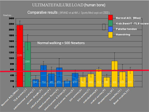
Strength of new technique (green) versus traditional methods:
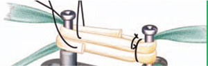
Preparation of 4 strand hamstring graft:
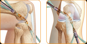
Position of graft as it is fixed in the knee:
Post-Operative Rehab
First 2 Weeks:
- Partial weight only on the operative leg with a knee brace
- Work on getting knee from fully straight to 90 degrees
- Start exercises in brace
Weeks 2 to 6:
- Put full weight on leg
- Get complete knee motion back
- Allow increased motion in brace for walking
- Progress exercises
Weeks 6 to 12:
- Work on strengthening
Week 12:
- Start running program
- Do objective strength test
12 weeks and on:
- Once reach strength goal begin sport specific activity
- Progress over next couple months to sporting activity based on strength testing and functional testing